Friday, January 30, 2009

Food Additive Combinations and ADHD

The concept of food additives, which include artificial colors and flavorings used in food processing, and their influence on ADD and ADHD is nothing new. Starting in the mid-1970's by Feingold as well as others, the idea that artificial food ingredients may have some type of pharmacological impact on neurodevelopmental disorders became a hot topic of discussion.

Today, the debate rages on as to how much of an effect these chemical ingredients really have on our systems. I am not going to lend my full support to either side of this discussion any time soon, because the evidence is strong for both arguments. Instead, I wanted to look at a less-discussed but equally important topic on the effects of food additives and ADHD, namely the synergistic effects of these compounds.

In terms of our discussion today, a synergistic effect is where two or more compounds or chemicals, when used in combination together, result in a greater impact than the sum of their individual effects (the concept of the "whole" being greater than the "sum of the parts"). For example, if a specific concentration of food chemical "A" reduces nerve cell growth by 10% and a specific concentration of food chemical "B" reduces growth by 15%, then, theoretically, a combination of these two concentrations together should decrease cell growth by about 25%. However, if the two chemicals combined (and all other factors being carefully controlled) reduce growth by, say 50%, then the cause is likely a synergistic effect or interaction between chemicals "A" and "B".

The investigation into synergistic effects of food additives stems from an article done by Lau and coworkers on how four food additives, well-known for their potential neurotoxic effects as individual agents, can potentially be even more devastating when used in combination.

The four food additives in question were as follows:

  1. Brilliant Blue, also referred to as "Blue1" and "E133" (in Europe)
  2. Quinoline Yellow, also referred to as Yellow 13 or E104
  3. Aspartame (Nutrasweet, Equal): and artificial sweetener often used in diet soft drinks
  4. MSG: short for Monosodium Glutamate or a salt form of L-glutamic acid, often used in Chinese foods and, (to a lesser extent now), potato chips and french fries

The study found that two pairings of the above compounds had notably significant synergistic effects. Brilliant Blue, when combined with MSG, showed a strong decrease in a process called neurite outgrowth. Neurite outgrowth, essentially, is the process where neurons begin to develop and differentiate, and eventually results in the interaction of neurons with either other neurons or cells of different systems such as muscle cells. In addition to the Brilliant Blue and MSG combination, the combination of Quinoline Yellow and Aspartame also showed a strong additive effect on inhibiting neurite outgrowth.

The process of neurite outgrowth is a major indicator of overall cell health with regards to the nervous system. Additionally, this process is especially critical during the neurodevelopmental stages, which starts during embryonic development, and can continue on until an individual is in his or her 20's. However, the period of greatest development (and greatest potential sensitivity to chemical agents), is between the sixth month of gestation to the first few years after birth. As a result, (in my humble opinion) anything that inhibits this process, should be taken seriously, especially during the early developmental stages in life.

It is also worth mentioning that the levels of these different chemical agents done in the study by Lau were below concentrations which typically cause neurotoxic problems on their own. In other words, these two combinations (Quinoline Yellow/Aspartame, as well as MSG/Brilliant Blue) showed extremely pronounced effects with regards to inhibiting key neurodevelopmental processes. Between these two combinations, the combined effects of Quinoline Yellow and Aspartame were more pronounced than the MSG/Brilliant Blue.

As far as the status of these four agents is concerned, three of the four (MSG, Brilliant Blue and Aspartame) are currently available in the United States, with Quinoline Yellow being banned. In the United Kingdom, where the study was done, all four of the compounds were still used in food processing. Brilliant Blue, while used in the US and UK, has been banned in most of Europe.

It is believed that the two flavor enhancers, aspartame and MSG both work via a type of biological receptor proteins called NMDA receptors. Without going into too much detail here (we will save the NMDA receptor topic for future posts), NMDA receptors play a huge role in the regulation of ion channels, which are critically important in a number of processes in a number of systems, including the nervous system. One of the key "target molecules" for these NMDA receptors is glutamate, which, as we've seen above is the major component of MSG. Additionally, part of the molecule of Aspartame is comprised of a form of aspartate, which is a form of a common natural dietary amino acid and is chemically similar to glutamate.

The reason that the above information is relevant to our topic of discussion is that glutamate and NMDA are both key biological agents involved in neuro-signaling processes which are significant factors with regards to ADHD and other disorders. In other words, chemical agents which interfere with this NMDA/glutamate "channel", often can, at least in theory, have an effect on the onset and symptomology of ADHD. We will go into much more detail on this process in later blog entries.

In addition to these concerns, we must also be aware of the fact that the NMDA receptor is a target of a number of different drugs and pharmacological agents. As a result, there is also the potential for synergistic effects between food additives and NMDA receptor drugs. In addition to current concerns of negative drug-drug (and now food additive-food additive) interactions, we must also be careful with regards to potential drug-food additive interactions. These interactions are easy to overlook, and, given the abundance of artificial food additives, are almost impossible to avoid completely.

Even if these four agents listed above all become banned at some point, I personally believe that this study should raise an alarm and open the way to a number of future studies on the effects of specific combinations of food additives. As highlighted in the article, one of the main problems with "elimination" diets for food allergies or toxicities, is that they often examine the food compounds in isolation, as opposed to combination. This study hopefully sheds some light on the fact that, perhaps, instead of just looking at individual food additives and their negative effects on ADHD and other neurodevelopmental disorders, we should be paying an equal amount of attention to investigating the negative effects of different combinations of these ingredients, especially the most common food-additive combinations that are currently available.

Add to Technorati Favorites

Monday, January 26, 2009

ADHD vs. OCD: Brain regions and bloodflow patterns

ADHD and OCD (obsessive compulsive disorder) are two disorders that often fall at the opposite end of the neurochemical spectrum. However, the two disorders, which are sometimes comorbid (occur alongside one another), actually share a fair degree of similarity with regards to underlying causes. It is believed that both disorders are the result of chemical imbalances in similar brain regions. One of these brain regions is the prefrontal cortex (orange region in figure below, please click here for original image source):


In the brain diagram above (side view, the left is the front of the head), the area highlighted in orange constitutes what is referred to as the prefrontal cortex region. We have previously alluded to the connection between the prefrontal cortex region and ADHD. It is believed that levels of the free signaling neurotransmitter dopamine are significantly lower in this region of the brain in ADHD individuals.

In addition, evidence strongly suggests a reduction in blood flow in the prefrontal cortex for ADHD individuals. This region can be likened to a "braking" region in the brain, in which inhibitory judgment and control of behaviors is thought to occur. Therefore, under activity in this critical brain region, either via deficiency in blood flow or chemical signaling agents and processes is thought to reduce the ability of the individual to inhibit unwanted responses and behaviors. As a result, impulsive behavior, which is a hallmark characteristic of ADHD is more likely to occur if this brain region is underactive.

On the other hand, this brain region has also been implicated as a critical brain region in cases of Obsessive Compulsive Disorder (OCD). However, unlike ADHD, where the prefrontal cortex region of the brain is believed to be underactive, in cases of OCD, the prefrontal cortex activity is thought to be overactive.

An increasingly popular method of determining brain activity is performed by using a process called SPECT. SPECT, which is short for Single Photon Emission Computed Tomography, utilizes a radioactive tracer of a compound which involves a radioactive isotope of the element Technetium. This chemical complex of Technetium can be used as a tracer to measure blood flow patterns to the brain, which is a method of detecting overall brain activity. In general, the higher the level of this "marked" blood, the more active a particular brain region is thought to be. This can be especially useful in determining which brain regions are used for specific tasks, such as problem solving or exercises involving heavy concentration. It is often the change in blood flow patterns between "resting" or "relaxed" states vs. cognitive tasks which can often give a clear picture of information as to how "hard" a specific brain region is working to complete the specific task.

While the above process is relatively safe and non-invasive, the idea of doing brain scans involving radioactive tracers and gamma radiation detection methods have significantly limited the use of this method of brain imaging in children and adolescents. However, since both ADHD and OCD often present themselves as disorders originating in early childhood or adolescence, relatively few studies have been done on SPECT-derived brain images in these individuals.

Returning to the ADHD vs. OCD topic of discussion, a relatively recent study was performed using children and adolescents with one of these two disorders, making it one of the few reported studies of its kind involving this particular age group of the population. Although relatively small in the number of test subjects, the study did confirm earlier presumptions regarding blood flow and brain activity of these two disorders, such as in a recent SPECT study on adults with ADHD.

The study found that there was a significantly lower level of blood flow (and therefore brain activity) in specific brain regions for the ADHD children vs. those seen in the OCD children. A strong attempt was made to compare images of ADHD vs. OCD children of the same age and gender in order to reduce the impact of developmental differences.

Below is rough sketch of some of the brain regions compared between the ADHD and OCD children from the study (for original image source, please click here). In this diagram, we are looking at the brain from the right side of an individual facing to the right. The term "cortex", used throughout this post, refers to the outer layers of a particular region. The term "prefrontal" cortex, as seen in the previous diagram, typically refers more toward the outer layer of the brain, right behind the forehead region:


Using the above diagram as a reference, here are some of the findings by Oner and coworkers regarding the differences in cerebral blood flow between ADHD and OCD children:


A quick word of caution: I am not going to go over the statistical methods used in the study in detail. However, given the relatively small sample size and numerical "cutoffs" for a difference to be statistically significant (as opposed to getting a difference just because of random chance due to natural variations with regards to sample sizes), the only region which met the criteria of being statistically significant in this study was the right prefrontal cortex. Nevertheless, there were some differences in blood flow patterns for some other brain regions, which, while not statistically "significant", were still somewhat noteworthy. The left prefrontal cortex should be noted in particular. Keep in mind that for this region it appears that activity is higher in ADHD than OCD, while the opposite is true for the right prefrontal cortex. I thought this difference was worth at least a mention in this post.


A few things worth noting from these differences in brain function between OCD and ADHD individuals:
  • Both ADHD and OCD are believed to be disorders associated with the glutamatergic system. While we will not go into too much detail here, glutamatergic activity involves glutamate, which is a form of one of the common amino acids and is a major neurotransmitting (a signaling process between cells in the nervous system) agent. ADHD is believed to be a hypoglutamatergic disorder (lower than normal activity of the glutamatergic system) while OCD is believed to be hyperglutamatergic (higher than normal activity of the glutamatergic system). In other words, ADHD and OCD are two disorders which are both believed to be imbalances of the same signaling or neurotransmitting system, but on opposite sides of the spectrum.

  • ADHD, OCD and Tourette's Syndrome may all share a common pathway involving a group of brain regions called the CSTC (which is short for cortico-striato-thalamo-cortical pathway). While I will not go into any more detail here, and save this for future discussion, this potential connection is worth mentioning because these three disorders often have a relatively high degree of overlap. We have already investigated some of the overlap between ADHD and Tourette's in previous posts.

***Please note: I do not want to open the door of erroneously linking multiple unrelated disorders together. I believe that it is one of the negative tendencies of researchers to attempt to link multiple disorders together based on insufficient evidence in an attempt to find some sort of unified underlying cause to everything. While I admit that I myself am susceptible to this natural bias as well, I try to avoid making these types of false conclusions as much as possible. Nevertheless, the last point was meant more to illustrate that a number of disorders which have been frequently listed as comorbid to ADHD do tend to exhibit differences in overlapping brain regions, especially the prefrontal cortex. In my opinion, the prefrontal cortex is, therefore, potentially the most critical brain region to study when investigating ADHD comorbid disorders.

While the prefrontal cortex region is a crucially important brain region with regards to ADHD and related disorders, it is by no means the only one involved in these processes. We will investigate some of these other key brain regions in posts in the near future.

Add to Technorati Favorites

Thursday, January 22, 2009

ADHD, Alcoholism and Nutrient Deficiencies

This will probably be the last blog post on the ADHD and alcoholism connection. We have investigated the connection between ADHD and alcoholism with regards to:

Now we will investigate another potential connection between the disorders of ADHD and alcoholism, which involves alcohol-induced deficiencies of key vitamins and minerals which are often deficient in individuals with ADHD. We will list some of these key nutrients below:


Magnesium: (Here are recommended daily magnesium intake levels)

We have posted on this nutrient extensively in the past. For example, there is relatively strong evidence of a connection between magnesium deficiency and childhood ADHD. Additionally, there are a number of disorders which occur alongside of ADHD, which are called comorbid disorders. Magnesium levels are thought to influence some of these ADHD comorbid disorders as well. Co-treatment with vitamin B6 has been shown to boost magnesium's effects for ADHD treatment as well. Finally, I have outlined some other nutrient treatment combinations thought to boost the effectiveness of magnesium for ADHD.

Magnesium deficiencies are also common in chronic alcoholics. There are several potential reasons for this including decreased absorption and increased urinary loss of magnesium, dietary deficiencies as alcohol calorically replaces magnesium-rich foods, and decreased retention due to liver dysfunction. Unfortunately, the actual process of quitting alcohol use can also result in magnesium shortages. This is due to the alcohol withdrawal process in which results in fatty acid composition changes and the buildup of compounds in a process called ketoacidosis. These compositional changes during the alcohol withdrawal process can result in products which bind to magnesium and reduce its serum levels. A review by Krishnel and coworkers on the efficacy of intravenous vitamins for alcoholics in the emergency department touted the benefits of oral magnesium supplementation for admitted alcoholic patients.


Thiamine (also spelled "thiamin"): (Here are recommended daily thiamin intake levels).

There are several studies pointing towards a connection between chronic alcohol abuse and thiamine deficiency, although the scope of these effects is still under debate. Thiamine deficiency has been implicated for a disorder called Wernicke's encephalopathy. Wernicke's encephalopathy does have some overlap in symptoms with ADHD, such as impaired short-term memory, but beyond this, there is little connection between the two disorders. One thing to note about thiamine is that while there is minimal research done on the possible connection between its deficiency and ADHD, thiamine does play a major role in the process of glucose metabolism. Individuals with ADHD have often shown sub-average blood glucose levels to several key brain regions. Some studies have even implicated a potential risk of thiamine depletion caused by rapid glucose administration (such as through IV treatment).


Vitamin B-6: (Here are recommended daily vitamin B-6 intake levels)

Vitamin B-6 has had numerous implications for both the causes and treatment of ADHD. B6 has been shown to assist and boost the effects of magnesium in treating ADHD. Vitamin B6 has an "active form", which is often referred to as pyridoxal phosphate (PLP).

Chronic alcoholism can lead to a condition known as hyperhomocysteinemia. This disorder is the result of excessive buildup of the compound homocysteine. Homocysteine has been implicated as a major factor in a number of cardiovascular and inflammatory diseases and is a leading culprit of stroke and arterial damage. In addition to these disorders, high homocysteine levels are thought to play an indirect role in the onset of ADHD.

Vitamin B-6, vitamin B-12 and folic acid all play a role in regulating homocysteine levels. In fact, there is thought to be a minimal level for each of vitamin B6, B12 and folate to combat excessive homocysteine levels. Below is a rough sketch of how homocysteine is converted to the more benign and extremely important bodily antioxidant glutathione. This is important, because ADHD individuals have often been shown to have lower than normal levels of this ubiquitous antioxidant (as well as antioxidant levels in general). Upping the conversion of homocysteine to glutathione through B vitamin-dependent pathways therefore presents two different therapeutic measures for the ADHD sufferer.


At this point, there is no need to familiarize yourself with the intermediate steps in the process, just note that the "active" form of vitamin B-6, Pyridoxal phosphate or PLP is needed in not one, but two different steps of this conversion process. Low levels of this key nutrient can lead to a backup of homocysteine as this process is severely hampered.

Vitamin B-12: (Here are recommended daily vitamin B-12 intake levels)


As mentioned above, vitamin B-12 also plays a critical role in maintaining homocysteine levels. It, along with folate (the "nutritionally active" form of folic acid), actually work together, along with a third compound called betaine) in converting potentially dangerously high levels of homocysteine back to the amino acid methionine. Keep in mind that deficiencies of vitamin B-12 can cause problems with regards to homocysteine buildup as an under balance of vitamin B12 with respect to folate can boost homocysteine levels. Keep this in mind when we proceed to the folic acid discussion, as isolated supplementation with folate can offset the desired B12/folate balance and be counterproductive. A brief diagram of this process can be seen below:

A quick note: If you look at the diagram above, you can see that the process of removing homocysteine by converting it to methionine can actually continue on to another important compound, S-Adenosylmethionine (SAMe). There has been a lot of discussion surrounding SAMe as a possible supplement used to treat ADHD. We will save this discussion for a later time, but it is at least worth mentioning that there have been some very positive things said about this nutrient. Additionally, SAMe has been shown to help protect against liver damage (even to the point of reversing the process), which, as we know, is extremely common in alcoholics. Also note that betaine supplementation can also help offset alcohol-induced liver damage, so the betaine mentioned in the above process is multifunctional with regards to ADHD and alcoholism.

In addition, there may be a connection between vitamin B-12 deficiencies and food allergies (which are often associated with a rise in ADHD-like behaviors themselves). This is in part, due to the connection between B-12 deficiencies and pernicious anemia. This is characterized by a reduction of gastric acid secretion through damage to cells in the stomach called parietal cells. Food allergies, which have been associated with ADHD, can be exacerbated by weak stomach acid levels, as food allergens which are normally broken down by sufficient acid are now present at higher levels. We have seen the effects of damage to the stomach and other digestive organs in the case of our earlier post on celiac disease and its correlation with ADHD symptoms.

***Keep in mind that this B-12/food allergy and ADHD connection is more hypothetical at this point, relatively little published information is available to confirm this indirect connection. Nevertheless, I personally believe that this possible association is at least worth mentioning.

Folic Acid/Folate: (Here are recommended daily folate intake levels)

As alluded to above, we have seen the intricate connection between vitamin B-12 and folate (folic acid is the synthetic form of folate used in food fortification. Within the scope of this post, I am using the two terms interchangeably). With regards to cognitive function and relevant disorders such as ADHD, there is also an important relationship regarding the balance of these two nutrients. For example, a relatively recent study found that for vitamin B-12 deficient individuals, folate is actually connected to folate and reduced cognitive function. However, when ample B-12 levels were available, higher folate levels were protective against cognitive impairment. Thus we see that folate can potentially be a double-edged sword in the war against high homocysteine levels and reduced cognitive function, and that folate's effectiveness is grossly dependent on an adequate vitamin B-12 balance.

Aside from the homocysteine/B-12 connection, it also appears that folate plays other critical roles which can indirectly affect the severity of negative symptoms associated with ADHD. Additionally, folic acid has been found to have a protective effect against formic acid, a neurotoxin. This relationship actually stems from the neurotoxic effects of methanol, which is often found in alcoholic beverages either as a congener (essentially a side product in alcoholic beverages, which actually play a factor in the hangover process), or through endogenous formation (within the body). One of the problems with methanol is that it shares the same enzyme system as ethanol (the main form of alcohol in beverages), but is slower to clear due to a less-efficient metabolic process and can build up to toxic levels in heavy drinkers. However, adequate folate levels in the liver can expedite the methanol metabolism and clearance and reduce levels of the neurotoxin formic acid. In addition to the liver, there is some evidence that folate-derived formic acid metabolism occurs in the mammalian brain as well. Folate is also thought to be connected to the key compound in regulating levels of SAMe (S-Adenosylmethionine). Folate deficiency can lead to reduced levels of SAMe. This is of importance, because in numerous studies S-Adenosylmethionine has been implicated as a potential treatment option for ADHD.


A quick word on homocysteine: We have spent a fair amount of time highlighting the connection between alcohol consumption and homocysteine levels. In fact, chronic alcoholics reported double the serum homocysteine levels as nondrinkers. Hyperhomocysteinemia has also been associated as a major culprit in the process of alcoholism-induced brain shrinkage.

However, it is worth noting that the source of the alcohol may play a critical role with regards to homocysteine levels. A study found that beer consumers had notably lower levels of homocysteine than did consumers of wine or other spirits. While this association was not thoroughly addressed, this is possibly due to the relatively high levels of B vitamins in certain forms of brewer's yeast (which is used in the beer-making process). This is right in line with our study on vitamins B-6 and B-12.

In addition to the nutrients listed above, there are thought to be other nutritional factors at play. For example, chronic alcoholics who are faced with alcohol withdrawal are at increased risk of omega-3 fatty acid oxidation. This oxidative damage can disrupt the omega-6/omega-3 fatty acid balance, which we addressed in an earlier post as being a critical factor in cell membrane integrity. Additionally, alcoholism has been linked to deficiencies in antioxidants such as vitamin C (remember that individuals with ADHD generally have lower total antioxidant levels than their non-ADHD peers). Alcoholic liver damage has also been linked to zinc deficiency. We have investigated the zinc connection to ADHD earlier, namely in the potential ability of zinc to boost the effectiveness of Ritalin, a common ADHD stimulant medication.

Finally, I have alluded a bit to the compound S-Adenosylmethionine (SAMe) in this post. It is an ADHD treatment method of great potential interest. We will be discussing the possible merits of SAMe in the near-future.

Add to Technorati Favorites

Friday, January 16, 2009

Genes, Omega-3's, Alcohol and ADHD

In our last discussion, we were exploring the theory behind omega-3 fatty acid supplementation for ADHD, and alluded to the fact that there may be some genes at work involving this process. Additionally, there is some evidence that alcohol use can inhibit the effectiveness of some of the enzymes that are coded for by these genes, and possibly be a factor in the onset of ADHD. We will be exploring these associations in this blog post.
Omega-3 fatty acids are crucial for our overall well being for a number of reasons, with many of them being tied to maintaining the structure of all different types of cells in our bodies. Among these omega-3's are alpha-linolenic acid (ALA), eicosapentaenoic acid (EPA), and docosahexaenoic acid (DHA). ALA converts to EPA (and eventually DHA) through a series of steps, several of which use the enzymes governed by the genes listed above. A summary of this process is highlighted below (original file source here):

The diagram above may look quite complicated, but we're just focusing on a few of the objects listed above.

As a quick side note: a lot of the other objects on this diagram above are showing the role these omega-3's and omega-6's play in the inflammatory process of immune reactions. This discussion is beyond the range of this post, but I have included it to illustrate that omega-3 and omega-6 fatty acids play a critical role in regulating a number of different functions and systems. Omega-3 imbalances can lead to immune dysfunction, which is thought to be one of the reasons why individuals with ADHD, who often have lower blood levels of omega-3's than their peers, are also more likely to have immune system disorders such as allergies. This ADHD/allergy connection will be explored in the future. Also, notice that omega-3's and omega-6's use the same enzymes. This is important, and was discussed at length in the previous post.

The section on the left of the above diagram describes how one omega-3 fatty acid is converted to another, for example, the a-linolenic acid (top left) eventually makes its way to forming EPA (fourth one down on the left), which eventually is converted to DHA (last one in the left column). Bringing our attention to the center, we see a series of enzymes with names like ∆6 desaturase, elongase, etc. These enzymes play a major role in the actual chemical conversion process of one type of omega-3 fatty acid to another.

Keep your attention on the enzymes that have the key term desaturase in their title. These are the ones we need to be concerned about when dealing with the aforementioned genes and alcohol. Without these enzymes functioning at their highest level, the incorporation of dietary omega-3's into the actual structure of the cell membrane is significantly. Genetic differences and the presence of external factors (such as alcohol or other types of fats) can significantly impair the function of these enzymes and slow the conversion process (and ultimately uptake and incorporation into cell membranes) of these critical omega-3's.


A number of these desaturase enzymes are all coded from a specific genetic region located on the 11th chromosome in humans, located at the 11q25 region (chromosomes have 2 "arms", a "p" and a "q", the numbering refers to relative location on that arm, so "11q25" refers to the 25th region on the "q" arm on the 11th chromosome). Interestingly, this region is located near the 11q22 region, which has been linked to ADHD. The closer two genetic regions are, the higher the chances they will be co-transmitted (passed on together from parent to child). In other words, gene forms which are located near each other on a chromosome are more likely to be passed on together, suggesting the possibility that the 11q22 ADHD region may in fact be influenced by some of the genes from nearby 11q25 region.

Brookes and coworkers did a study on the association between these desaturase genes and ADHD (on a personal note, I would like to acknowledge the authors of this particular study. Much of the information in these past two posts is gleaned from their work, and this paper provided a great starting point for much of my research for this post). They found that the 11q25 region contained three genes which code for desaturase enzymes located next to each other: Fatty Acid Desaturase 1, Fatty Acid Desaturase 2, and Fatty Acid Desaturase 3 (abbreviated as FADS1, FADS2 and FADS3, respectively). These genes each exist in different forms, called alleles, which have slightly different DNA configurations (which can differ by as little as one letter in the DNA "code").

Key findings from the Brookes study: This group saw a significant difference in the prevalence of ADHD stemming from two different alleles in the FADS2 gene. It appears that a single point difference was all it took to boost the likelihood of association with ADHD. Individuals with ADHD were significantly more likely to have the "C" form of the FADS2 gene than the "T" form of the gene at marker 498793 (this number just gives the detailed location on which spot of the DNA this form can be found).

Additionally, it appears that the onset of ADHD stemming from prenatal alcohol exposure may be somewhat genetic as well. For individuals who were exposed to alcohol via maternal consumption during pregnancy, there is some nominal evidence linking "G" allele instead of the "C" allele at two different locations on the FADS1 gene was correlated with a higher likelihood of being diagnosed with ADHD. However, the authors concluded that this connection was only "speculative".

This possible ADHD/genetics/fatty acid consumption/alcohol exposure connection is somewhat intriguing. The study established a strong ADHD connection to a specific allele of the FADS2 gene on the 11th chromosome, and also cited a number of other studies on the effects of omega-3 consumption on ADHD symptoms, but the connections with alcohol are more strained. Nevertheless, the findings from other studies offer support for this possible alcohol association with these other factors:
  1. We have seen before that omega-3 fatty acid deficiencies are more prevalent in individuals with ADHD. The previous post describes the process of how omega-3's affect cell membrane integrity, which, in turn, can effect the passage of key chemical signaling agents such as dopamine (which has repeatedly been found to deficient in specific brain regions of ADHD individuals). The desaturase enzymes, which are products of the genes listed above are partly responsible for the process of omega-3 metabolism and incorporation into the cell membranes.


  2. Different alleles (alternate forms of a gene) can result in slightly different forms of these enzymes, some of which are more efficient than others. In other words, enzymes coded for by one form of a gene are somewhat better at metabolizing omega-3's and incorporating them into cells than the "alternate" enzymes coded for from the "alternate" forms of the gene. As a result, small changes in the gene code in these aforementioned regions can indirectly affect the efficiency of omega-3-to-membrane incorporation.


  3. Several studies have pointed to the the connection between alcohol and fatty acid metabolism in animal models of ADHD.


  4. It also appears that an individual may be able to "recover" from some of the negative effects on cognition due to alcohol exposure by an increase in dietary omega-3's. This includes increasing maternal dietary levels of omega-3's during pregnancy (based on animal model studies).

To summarize the whole post (as well as the previous one), it appears that omega-3 fatty acid metabolism plays a major role in ADHD. This is thought to be at least in part to the effects of omega-3's on maintaining cell membrane structure and integrity and their effects on regulating levels of the brain signaling agent dopamine (which is a crucial neurotransmitter and is often deficient in ADHD cases). However, properly functioning enzymes are required for these steps. Desaturase enzymes are coded for by a genetically "hot" region for ADHD on the 11th chromosome in humans. Different versions of these genes can result in a reduction in enzyme function and potentially affect the way these omega-3's are metabolized. In mammals, alcohol exposure can also lead to reduced desaturase enzyme activity. Additionally, there is at least some evidence that alcohol can increase the likelihood of specific forms of FADS1 gene giving rise to ADHD. This may be due to the two factors combining to reduce desaturase enzyme activity to a point where omega-3 metabolism falls past a hypothetical "break-point" resulting in a sharp increase in the onset of ADHD and other related disorders.

We have been focusing heavily on the ADHD and alcoholism connection for the past couple of weeks. We will be investigating a few more studies on this connection in the upcoming posts.

Add to Technorati Favorites

Monday, January 12, 2009

Omega 3 Fatty Acids and ADHD: The Theory Behind the Practice

How Omega 3 Fatty Acids work and their influence on ADHD

In the past couple of posts, we have examined the connections between ADHD and alcoholism. We will continue this discussion shortly, when we begin to investigate specific genes of overlap between the two. One of these genes, whose products are thought to be affected by alcohol consumption, and appears to have some degree of influence on ADHD is called the Fatty Acid Desaturase 2 gene. We will be investigating this gene in the next post, but I want to preface it with a bit of a background information as to why fatty acids, especially the famous omega 3's, are believed to be so attractive as potential natural treatments for managing ADHD (as well as a host of other disorders).

Since ADHD is so strongly affiliated with the nervous system, the physical composition of this system is extremely important when considering some of the implications for this order. Keep in mind that the brain is over 60% fat in humans and other mammals.


Additionally, during the brain developmental stages, neurons are coated with an insulation of sorts, a fatty material called myelin. This whole process is called myelination. When this myelination process is complete, a neural connection can be up to 100's of times more efficient, and signaling through these connections can become exponentially faster. During the teen years, this myelination process often runs rampant, as the brain begins to hardwire itself for greater efficiency. That is why it is so crucial to develop these key connections early in life, before this myelination process begins.

Given the importance of fat in the myelination process, and the overall abundance of fat in the brain as a whole, the nervous system is extremely influenced by fat composition obtained from dietary means. Cell membranes, which are the outer protective layers of cells (in all parts of the body) are also comprised of fatty materials. Among these are omega-3 fatty acids and omega-6 fatty acids.

**Please note: the rest of this post deals primarily with the biochemistry of omega-3 fatty acids and their impact on cell structure and function, and their connection to disorders like ADHD. If you are just interested in general strategies on omega-3 supplementation, you can skip to the end of the post, where I have listed 6 tips to increase your chances of effective treatments. If you want a bit more background as to why I am giving these suggestions, please continue reading!

These two types of fatty acids each have unique structures, which means that their incorporation into the cell membrane also affects its structure. For example, omega 3's typically take on a more curvy shape, and omega 6's are often more "straight" and narrow. Because of these shape differences, the omega-3 rich regions of the cell membrane are more prone to forming "gaps" in the cell membrane, making this whole region more "fluid". However, the straighter, more rigid, omega-6 regions of cell membranes make for tighter and smaller gaps, making the cell membrane less flexible. Numerous studies have shown that fatty acid composition in cell membranes is directly affected by dietary intake of omega-3 fatty acids.


Among the omega 3-fatty acids, perhaps the most important is called alpha-linolenic acid (ALA). The human body is unable to produce this type of fat, so it must be obtained via dietary measures. The body can then convert ALA to two other types of omega-3 fatty acids, DHA (docosahexaenoic acid) and EPA (eicosapentaenoic acid). Both DHA and EPA can be incorporated into cell membranes, giving them a more flexible conformation. Not surprisingly, all three of these omega-3's (ALA, DHA and EPA) are currently popular supplements and health-food items. Supplementation with EPA and DHA-rich fish oil has been shown to boost levels of these omega-3's in the cell membrane.

Keep in mind that many of these studies of omega-3 incorporation into cell membranes typically involve blood cells as opposed to nerve cells. However, there have been a few recent findings supporting the incorporation of supplemented DHA into neuronal cells in mammalian systems. Additionally, dietary differences in omega-3 fatty acids has also been shown to influence the ratio of these to other fats in the brain in rat model studies of ADHD, and possibly influence learning behaviors.

The makeup and rigidity of the cell membrane is very important for proper functioning among cells in the nervous system. Gaps, such as those from omega 3 fatty acid regions, allow easier passage of key materials in and out of cells. Among these key openings are a type of passageway, made up of protein-based structures called ion channels. We will see in later posts that ion channels play a huge role in a number of diseases and disorders, including those which involve the nervous system (including ADHD). It is believed that these ion channels are not directly influenced by omega 3's and other fatty acids but rather by the tension on the cell membrane caused by these fats. Therefore, the right amount of tension, governed by the fatty acid composition is thought to regulate ion channel function is necessary for proper cell function.


Additionally, these ion channels are able to change shape, allowing the membranes of different cells to "fuse together" at specified times. This allows for adequate conductance of electrical signals and facilitates communication in-between cells. However, with a more rigid structure (i.e. from one that is deficient in omega 3 fatty acids), this lack of flexibility impairs the ability of these ion channels to change to the optimal conformations necessary for this fusion process. As a result, functional cell-cell communication is hampered. This too, is thought to be a factor in disorders such as ADHD (which will be discussed in future posts).

Perhaps the biggest effect that cell membrane integrity has to do with ADHD is its influence on the signaling agent dopamine. It has repeatedly been shown that ADHD is intricately connected to dopamine-based signaling methods and systems. The role of dopamine on ADHD is especially pronounced in specific brain regions such as the prefrontal cortex, in which this key neurotransmitter is often deficient. Numerous animal studies have shown that a deficiency of omega-3 fatty acids can lead to reduced dopamine function in the prefrontal cortex.

Interestingly, there has been a reported increase in dopamine levels in omega-3 deficient animals in another brain region called the nucleus accumbens. The reason this is somewhat intriguing is that the prefrontal cortex and the nucleus accumbens are thought to work in different directions, in an oppositional sort of way. Some studies suggest that this "ADHD" brain region, the prefrontal cortex inhibits the nucleus accumbens. As a result, a dopamine deficiency in the prefrontal cortex could lead to less inhibition and higher dopamine levels in the nucleus accumbens brain region. This confers the idea that the prefrontal cortex is often deficient in free levels of the important neurotransmitter dopamine.


When addressing ways to "naturally" treat deficits with regards to any type of disease or disorder, it is often tempting to "supplement" the problem away. Because of the dopamine deficiency in the prefrontal cortex, combined with the fact that omega-3 fatty acid deficiencies have repeatedly been seen in ADHD brains, it is easy to jump to the conclusion that rampant supplementation with fish oils and other omega-3 rich sources can make negative symptoms of this disorder go away.

However, research has indicated that although individuals with ADHD have been shown to have plasma deficiencies of omega-3 fatty acids, the cause is not likely to be a dietary omega-3 deficiency. Only a few limited studies have actually suggested direct reduction of ADHD symptoms with omega-3 fatty acid supplementation. For example, based changes in teacher rating scores on ADHD symptoms, children who took EPA and DHA supplements did show noticeable reductions in ADHD symptoms. Interestingly, this same study found that the effects of antioxidant vitamin E were also a large factor.

Even if these studies above hold true for the general population, numerous others have shown omega-3 supplementation to be effective in reducing ADHD symptoms. What is confusing is that this method has proven successful in some instances, while doing little-to-nothing in other cases. As a result, we are left with the big question, why? It appears that the answer may lie in the genes of the individual.

Fatty acid desaturase genes are responsible for coding for a series of enzymes of the same name. These fatty acid desaturase enzymes are important for the metabolism of omega-3 fatty acids. Deficiencies in fatty acid desaturase enzymes are not limited exclusively to genes. We now know that external chemical factors such as maternal alcohol use can also reduce the activities of these key enzyme systems. As a result, omega-3 metabolism suffers. Our next post will deal almost exclusively with this topic.

Before we go, I would like to list a few strategies to follow if you're interested in exploring omega-3 fatty acids as a treatment option for ADHD. Of course there is no guarantee that this treatment method will work, but here are a few pointers to stack the deck in your favor:

***Please note: You may be wondering why I am not giving specific dosage recommendations for omega-3's. There are two main reasons: 1.) There are still no clear-cut established daily amounts, and with the information I currently have, I am not fully comfortable in recommending a numerical amount, and 2.) Due to so many other factors at work (such as age, gender, disease status, cardiovascular health, genetic background, total caloric intake, and other dietary choices), omega-3 recommendations do not follow a one-size-fits-all model. However, a better option is to keep a good balance between omega-3 levels and intake levels of other fats. Since dietary fat intake plays a huge role on hormonal functions, overall ratios and balance play as much of a role as total amounts. Nevertheless, if you're looking for a rough estimate, many of the sources out there generally suggest levels of around 1-2 grams (on the higher end of this for men and the lower end for women) total of omega-3 fatty acids per day.

  1. Take a mixture of omega-3 fatty acids, not just one kind. Since ALA is the omega-3 precursor (mentioned above) to EFA and DHA, it might be tempting to just take ALA and let it convert to these other omega-3's in the body. However, this conversion process is slow and inefficient, as the enzyme system involved results in less than 1% of the ALA being converted to EPA and even less (since EPA goes through a series of steps using other enzymes to convert itself to DHA) to DHA.
  2. Don't omega-3 overload. This is extremely important. Many well-meaning treatment methods for ADHD and related disorders often try to force down high levels of these seemingly benign substances to "cure" these disorders. However, an omega-3 overdose can also cause problems. These enzymes (which are the same desaturase enzymes will will be discussing in the next post), operate by a mechanism called negative feedback. This means that if omega-3 levels are too high, the activity of these enzymes is significantly reduced, and the conversion processes listed in suggestion #1 are greatly impaired.
  3. On the other hand, keep a good balance between omega-3 fatty acids and omega-6 fatty acids. Recommendations may vary, but most sources recommend between a 1:1 and 2:1 ratio of omega-6's to omega-3's. Unfortunately, most Western industrialized diets have a much more skewed ratio, often upwards of 10:1 or even 50:1 in favor of the omega-6's. This imbalance, too, will affect enzyme activity in the omega-3 conversion process. As mentioned above, a balance of these dietary fats is essential for maintaining proper structure and integrity of cell membranes. While this is a bit of oversimplification, fats from marine sources are typically much greater in omega-3's and fats from land animals is higher in omega-6's (and another class of fats called omega-9's, which the body can actually produce from the other 2).
  4. Keep your vitamin E levels up to speed. Since the brain is comprised of high levels of fat, it is one of the most oxidation-prone organs in the entire body. A number of neurodegenerative diseases such as Alzheimer's are thought to be products of this oxidation process. While all antioxidants have some benefits, vitamin E appears to be one of the best with regards to brain health. This is in part because it is a fat-soluble vitamin (unlike vitamin C, which, in its most common form, is not). I mentioned in the study above on a reduction of ADHD symptoms based on teacher evaluations after omega-3 supplementation that vitamin E levels were also a major factor in the study.
  5. On the other hand, don't go overboard on the vitamin E. General daily amount recommendations and upper limits (a bit high in my opinion for the upper limits, try to stay well under these upper boundaries), and food and supplement sources of vitamin E can be found here. While a number of antioxidants are water-soluble, like vitamin C (which can easily be flushed out of the system and much tougher to overdose on), vitamin E can build up to toxic levels in the body much more easily. An alternative strategy is to take sufficient levels of vitamin C, which can help "recycle" vitamin E and enhance it's positive antioxidant effects while reducing the likelihood of toxicity.
  6. This should go without saying, but eliminate alcohol intake if you are pregnant. We will spend our next entire post on the negative effects of maternal alcohol consumption on these desaturase enzymes which are needed to convert dietary omega-3's to ones which can be used by the cells. This is another possible link between alcohol and ADHD, a topic which we have been exploring in quite a bit of depth as of late.

Add to Technorati Favorites

Saturday, January 10, 2009

ADHD: A Precursor to Alcoholism? (Corpus Callosum part 2)

In the previous blog post on ADHD and alcoholism, we investigated some of the signs of overlap between the two disorders. One factor which both disorders appear to share in common is a reduction in volume of specific brain regions, including the corpus callosum, the genu, and the isthmus, when compared to similar individuals who do not exhibit either of the symptoms of ADHD or alcoholism. For a generalized location of these brain regions, please see the diagram below (which is a reprint of the figure from the last blog entry):

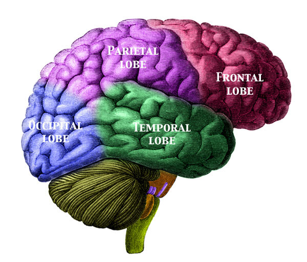
The corpus callosum is the tan band located inside of the gyrus cinguli, also known as the cingulate gyrus, a brain region whose role in ADHD we have discussed previously. The isthmus region is relatively small and is not labeled on this diagram. It is located to the right of the label "corpus callosum", and is just to the left of the area labeled "splenium".

**Please note my use of nomenclature here, I am continuing to use the same labeling as I did in the previous post. Here, we are considering the "genu" and "isthmus" as separate, distinct regions from the corpus callosum due, in part, to how they were classified in the studies I have been reviewing. However, many professionals label them all as one entity, with the genu and isthmus serving as sub-sections of the corpus callosum as a whole.

In today's blog, we will discuss the answers to two key questions:
  1. Is there a hereditary factor at play for the size abnormalities for these three brain regions?

  2. Does the outward expression of symptoms for one of these two disorders predate the other? In other words, is the appearance of ADHD symptoms a warning sign of impending alcohol abuse (or vice versa)?

For the answer to the first question, we turn to a journal article by Venkatasubramanian and coworkers in the journal Psychiatry Research: Neuroimaging. This group found that the relative size of the corpus callosum, genu and isthmus regions of the brain were correlated to an individual being at "high-risk" or "low-risk" for alcohol abuse. In this study, the "high-risk" group had a statistically significant reduction in size of multiple brain regions when compared to the "low-risk" group.

For comparison purposes, the "high-risk" group had approximately:

  • a 9% reduction in corpus callosum volume
  • a 13% reduction in the volume of the genu, and
  • a 17% reduction in isthmus volume when compared to the low-risk group for these brain regions.

**Note: it worth mentioning that the comparative differences in relative brain sizes followed some sort of age-dependent trend. The subjects of study were boys and young men between the ages of 9 and 23. For the age 9-15 group, all three of the above brain regions were smaller. However, for the older group (over 15), only the isthmus region had a statistical size difference between the "high-risk" and "low-risk" groups. This suggests that either the isthmus region follows a much greater developmental delay in individuals identified as being "high-risk" for alcohol dependence than do the corpus callosum or the isthmus region never does fully "catch up" in size to the "low-risk group"

So what exactly constitutes "high" and "low" risk? In this study, the researchers studied males between the ages of 9 and 23 (sampling a relatively even distribution across this age range) who had male parents who both:

  1. Developed an alcohol dependence before the age of 25 (average age of dependence around 20)
  2. At least two relatives (first-degree), who also had alcohol dependence (average was 3 relatives).

The "low-risk" control group of children and young adults consisted of individuals whose parents were not diagnosed with alcoholism or any other psychological disorders. To ensure that maternal genetic influences were not a factor, none of the mothers of the children (the 9 to 23 year-old group) in the study were diagnosed with alcohol abuse.

This study is of potential interest. While the sample size was small (only 20 "high-risk" and 20 "low-risk"), the choice of following only male parents and male children offered some clear-cut advantages over other similar studies. Among them were:

  • It eliminated gender effects. While debatable, some studies have found brain regions like the corpus callosum to be larger in males than in females. Having an all-male study eliminated this potential factor.

  • The ages of the two groups (high and low risk) followed similar distribution patterns, to rule out size increases in these brain regions due to aging.

  • Since the alcoholism was restricted to the paternal side, physiological effects during pregnancy were eliminated. This is important, as several studies have shown that maternal alcohol consumption during pregnancy can affect the development process including relative sizes of these brain regions. In other words, confounding effects, such as fetal alcohol syndrome were significantly reduced, since none of the mothers in the study suffered from clinical alcohol abuse.

  • None of the 9 to 23 year-old test subjects had already developed an alcohol dependence. This eliminates the effects that chronic alcoholism has on reducing the size of the corpus callosum, as reported in recent studies.

  • Other than alcoholism for fathers of the "high risk" group, none of the parents of the study participants had any other psychological disorder. This is important, especially due to the effects of comorbidity (disorders or symptoms occurring alongside alcoholism, such as conduct disorders or abuse of other substances) on worsening the symptoms of alcoholism.

So how does ADHD tie in to all of this?

Based on this study's findings, it appears that the expression of ADHD (as well as other "externalizing symptoms", which are symptoms that can be seen outwardly, such as hyper-excitability, conduct disorders, substance abuse, etc.) may be a warning sign of impending alcohol abuse.

For example, it has been postulated that hyperexcitability, which is often seen in individuals with ADHD (especially of the hyperactive-impulsive or combined subtypes) is a genetic precursor to alcohol dependence. In this case, and ADHD-like trait predates alcohol abuse. In other cases, symptoms such as substance abuse, conduct disorders, impulse control problems and other antisocial behaviors have grouped under the umbrella term generalized disinhibitory complex. Here, these numerous symptoms are essentially "clumped together" as one generalized behavior.

Regardless of the "model" one subscribes to, please keep in mind that a reduction in size the corpus callosum, genu and isthmus regions of the brain have been associated with ADHD.

Additionally, the degree of overlap between the two disorders was definitely worth mentioning. Out of the 20 children and young adults of the "high-risk" group, 17 of them were diagnosed with ADHD. Out of the 20 "low-risk" children? Zero.

Keep in mind that the fathers of the high-risk group were not diagnosed with any other disorders, just alcoholism. Additionally, keep in mind that none of the high-risk children had developed an alcohol dependence as of yet. These facts give compelling evidence that fathers who suffer from early-onset alcohol dependence who are not even ADHD themselves, are much more likely to have male children with ADHD, with an onset which precedes alcohol dependence itself.

In essence, this study established a degree of linkage (but not necessarily a direct cause) between the expressed behaviors of ADHD and alcoholism and the physiological features of relative sizes of the isthmus, corpus callosum and genu.

Hopefully this provides evidence that there is in fact a strong underlying hereditary component surrounding both disorders of ADHD and alcoholism. In the next couple of posts, we will investigate some of the individual genes thought to be involved in both of these disorders.

Add to Technorati Favorites

Friday, January 9, 2009

ADHD and Alcoholism: The Corpus Callosum (part 1)

In our last post, we discussed some of the ties between ADHD and eating disorders such as bulimia. In this post, we will begin the first of a multi-part investigation on the connection between ADHD and alcoholism. In this session, we will see how these two disorders are both tied to improper function in a key brain region known as the corpus callosum.
Note the relative position of the corpus callosum in the diagram below (source of image here):


A quick aside: Note the proximity of this corpus callosum brain region to the cingulate gyrus (labeled "Gyrus cinguli" in the diagram above), a region which we discussed in a recent post on attentional control. The cingulate region can be thought of as the brain's "gear shifter". If underactive, it leads to consistent lack of focus on one thought or task (a hallmark characteristic of ADHD), if overactive, the cingulate can result in overfocus (a characteristic of obsessive compulsive disorder, or OCD).

Returning to the corpus callosum area of the brain, which is layered inside the cingulate gyrus, we can see some sub-regions of note. These include the genu and the splenium. There is also a small region (not listed on the diagram above), called the isthmus, which is just to the left of the splenium. Of these regions, pay close attention to the isthmus, genu, and corpus callosum.

Note: the classification of these brain region sometimes varies, some methods classify the isthmus and genu as part of the corpus callosum, while others group them as seperate elements. No need to get any further into specifics, but when I refer to "corpus callosum" in the context of this post, I am referring to the region distinct from the isthmus, genu and splenium.

The corpus callosum is primarily responsible for connecting and integrating information from the left and right hemispheres of the brain. It is composed of millions of individual fibers and is necessary for the integration and processing of sensory information and expressing this information through verbal language. This is one of the later-developing regions of the brain, and continues to develop and become more efficient during adolescence (and even into early adulthood). Studies have shown that this prolonged developmental process leaves brain regions like the corpus callosum more prone to improper development. One of the reasons young children have trouble expressing visual images verbally is because speech control is typically on the left side of the brain and visual imaging and imagination is typically on the right.

Improper development of this corpus callosum region can lead to quirks such as split brain. Additionally, it has been reported that development of this brain region can be impeded by prenatal alcohol exposure and is entirely missing in around seven percent of children with fetal alcohol syndrome. Additionally, chronic alcohol abuse can result in thinning in the corpus callosum region.

In addition to the inhibition of this key brain region due to alcohol exposure listed above, it appears that there may be an underlying factor at play for this region for both ADHD and alcoholism. A reduction in size in the corpus callosum, genu and isthmus has been associated with ADHD in both children and adults. A study done by Venkatasubramanian and coworkers found a connection between smaller volumes in these same three regions of the brain and an increased risk of developing alcoholism.

Note that a reduction in size of the corpus callosum has been linked with a decreased functional ability in this region as well. This includes the processing of information between the left and right hemispheres of the brain in processes such as integrating information of visual images obtained from both eyes.

In addition to its role in expressing and processing ideas and thoughts from both sides of the brain, the corpus callosum is also integral in coordinating movements in different parts of the body. This includes governing motor inhibition (restricting unwanted or inappropriate movements) across the body. Interestingly, individuals with ADHD have been shown to have a decreased ability in utilizing the corpus callosum to control movements, which is often tied to the impulsive behavior of ADHD individuals with their actions (such as constantly grabbing or playing with objects at inappropriate times).

The corpus callosum is not the only brain region thought to be involved with both ADHD and alcoholism. For example, the prefrontal cortex (the brain region behind the forehead), which we have discussed extensively in other posts, has repeatedly been found to be underactive for individuals with ADHD. Additionally, Schweinsburg and coworkers found a decrease in activation of the prefrontal cortex correlates with a higher risk in suffering from alcoholism.

In the next few posts, we will examine some of the genes thought to be underlying factors in both ADHD and alcohol abuse. Additionally, we will examine some of the numbers to get a better understanding of the magnitude of overlap between the two disorders. Finally, we will examine some of the "warning sign" behaviors which youngsters might display before the onset of alcoholism occurs.

However, in the next entry, we will examine whether there is a hereditary factor in place surrounding brain volume, as well the prevalence of expressed outward symptoms of ADHD, and how these are both associated with an increased risk in developing alcoholism later in life.

Add to Technorati Favorites

Tuesday, January 6, 2009

The ADHD and Bulimia Connection

ADHD is a disorder that has numerous comorbids ("comorbids" refer to disorders that often accompany or are seen alongside of ADHD). These include, but are not limited to: Depression, Tourette's, Conduct Disorders, Sleep Disturbances, Restless Legs Syndrome, Body mass and obesity issues, dysgraphia (poor writing skills and abilities), processing disorders, sensory integration disorders as well as several others.

In the midst of all of these co-occurring disorders, there are a few that often evade the attention of both researchers and the general public. One of these is the disorder bulimia nervosa. Bulimia nervosa (which is often simply referred to as bulimia), which is often characterized by eating (and often binging) followed by purging, is a major issue in many industrialized nations, especially among teens and young women. Based on a study by Surman and co-workers, it appears that there is a relatively high correlation and prevalence of bulima and ADHD. A link to a quick synopsis of the study can be found here, but for sake of time, I will summarize a few key findings from the article:

  • Impulsive behavior is a hallmark characteristic of ADHD, and impulsivity is also thought to be a major factor in bulimia as well. It is even hypothesized that some type of underlying factor may be responsible for governing both disorders.

  • Given the fact that the disorder of bulimia is expressed at much higher frequencies in young females in late adolescence and early adulthood, it is interesting to note that correlations between the two disorders were relatively weak for men and non-adult women. Additionally, this is worth mentioning because the percentage of individuals with ADHD is heavily skewed towards the male side. That being said, the fact that there was not more of a correlation between ADHD and bulimia in males could be a reflection of either a poor sample size or representation of t he general population, or a relatively weak connection between the two disorders (i.e., one this is unable to override the so-called gender bias of bulimia favoring women and ADHD favoring men).

  • These results were tallied from 4 relatively large sample pools previously constructed to evaluate the effects of ADHD over an extended, longitudinal, multi-year period of time. This suggests that some of these relatively strong bulimia/ADHD correlations did not appear simply due to random statistical chance.

  • Stimulant medications, such as methylphenidate, which are often the first line of treatment for individuals with ADHD, especially those showing pronounced signs of impulsivity and hyperactivity, have shown potential in the treatment of bulimia, albeit through studies with very small sample sizes.

Taking this one step further, it appears that genetics may be an additional overlapping factor involved in stimulant medication treatment for ADHD. For example, some research suggests that different forms of DAT1 may be responsible for the effects of methylphenidate on appetite and eating behaviors including purging (DAT is short for "Dopamine Transporter Gene"). We have seen previously that there is a connection between the DAT gene and ADHD. Located on human chromosome #5, DAT1 has been linked to Parkinson's, Tourette's and substance abuse.

Additionally, proteins coded for by the DAT gene are expressed in high concentrations in the basal ganglia region of the brain. The basal ganglia is essentially responsible, among other things, for determining how fast a person's brain "idles" For example, "type A" individuals, who are often workaholics, easily stressed, and always on the go at 100 miles per hour often have overactive basal ganglia, while the more relaxed, easy-going, "type B" personalities typically have less activity in this critical brain region. While there also appears to be a significant overlap between bulimia and depression, individuals with bulimia typically display higher basal ganglia activities than those with isolated depressive symptoms.

Given the prevalent distribution of this gene's expressed proteins in key brain regions like the basal ganglia, and the role of involvement of these brain regions in eating disorders, the DAT gene may be an important determining and regulating factor for bulimia and other eating disorders, especially in the context of comorbid ADHD.

Please note: These final remarks are simply this blogger's opinion on the subject:
I personally find this connection between ADHD and bulimia to be interesting. However, I do believe that we should be cautious when investigating ADHD comorbid disorders. It is tempting sometimes to fall into the trap of falsely assuming that correlation always implies causation, and trying to find underlying causes for disorders and attempting to link ADHD to every other disorder under the sun.

However, the role of the DAT genes, which have been tied to ADHD, do offer at least some credence to at least some degree of genetic predisposition to both ADHD and bulimia. This claim is further strengthened by the degree of overlap involving medication treatments of the two disorders, namely stimulants. However, there have been several documented cases of the disappearance of bulimia symptoms following treatment with methylphenidate (Ritalin, Concerta, Daytrana, etc.) for comorbid ADHD.

As a result, we may be faced with a "chicken and egg" question: "Does bulimia increase the risk of ADHD or does ADHD increase the risk of bulimia?" (or even "Are they both side effects of an even larger underlying cause?"). Another plausible explanation is that ADHD is a culmination of secondary effects involving bulimia and other eating disorders. Constant purging will typically wreak havoc on the digestive system and lead to improper food and nutrient absorption. I have hinted in previous posts that digestive disorders such as celiac disease can often manifest symptoms which closely approximate those of ADHD. Given the mounting evidence connecting ADHD (or other disorders which exhibit closely related symptoms which could potentially lead to a "false" diagnosis of ADHD if one is not careful) to nutrient deficiencies, it is quite possible that ADHD and its symptoms are secondary effects of nutritional deficits caused by eating disorders such as bulimia.

Add to Technorati Favorites